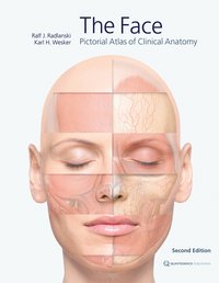
For the first time, the highly complex topographic-anatomical relationships of facial anatomy are depicted layer by layer using extremely detailed anatomical illustrations with a three-dimensional aspect. Important landmarks, anatomical details, and clinically relevant constellations of hard and soft tissues, as well as of nerves and vessels, have been detailed. Another important feature is that the point of view is maintained throughout while moving through the different layers of preparation. While the accompanying text and figure captions highlight specific issues, the images remain in the foreground. The elaborate illustrations are based mainly on live anatomy and corresponding images obtained from magnetic resonance imaging, with some support from anatomical preparations.